Viral disease of redgram
Pigeonpea sterility mosaic
Introduction
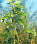
- This is a major disease of Pigeonpea in Karnataka. The district average incidence is highest in Bidar (49.70%) followed by Belgaum and Chitradurga.
- The characteristic symptoms of the disease are reduced growth, bushy and pale appearance, mild mottle and ring spot on leaflets. The plants may be wholly or partially sterile. The pathogen is transmitted by an eriophyd mite Acaria cajani Channabasavanna.
Methods of control or Management of virus diseases
Elimination of the source of infection
- Eradiction of weeds and other alternative hosts
- Roguing within the crop.
- Eradication of volumeteer plants.
Avoidance of the source of infection
- Modification of cropping procedures
- Cultivation in isolated areas
- Crop hygiene
- Use of virus free seed
- Use of virus free planting material
Avoidance of the vector
- Cropping in vector free areas
- Changes in cropping practices
Chemical control of vectors
- Air borne insect vectors.
Non chemical control of insect vectors
- Barrial and reflective mulches
- Biological control by predated
Control by cross protection
Control through resistant cultivars
- Host resistance to viruses
- Host resistance to the vector
Production of virus free plants
- Thermotherapy
- Virus eradication by tissue culture
- Chemotherapy
 Top Top
Alternaria leaf spot [Alternaria tenuissim (Kunze ex. Pers.) Wiltshire]
- This leaf spot disease is reported only from India where A.alternata has also been reported to cause a similar leaf spot. Initially small necrotic spots appear on the leaves, and these gradually increase in size to characteristic lesions with dark and light brown concentric rings with a wary outline and purple margin. As infection progresses, the lesions enlarge and coalesce.
- The disease is mostly confined to older leaves in adult plants, but may infect new leaves of young plants, particularly in the postrainy season crop. The pathogen is presented in the environment and has a wide host range. It was detected on pigeonpea seeds in Puerto rico. Resistant cultivars such as ICPL 366 and DA 2 are now available.
Top
Redgram blight : Phytopthora drechsleri f. Sp. Cajani
- This disease although not common in Karnataka, could become severe in infested waterlogged patches.
Symptoms
- Appearance of dark brown to purple lesions on stems, petioles and leaves, many a times resulting in death of the affected plants.
- Large number of resistant lines is available in the germplasm
- collections Ex., ICP 28, ICP 114, ICP 231, etc.
Top
Bacterial leaf spot and stem canker : Xanthomonas campestris cv. Cajani (Kulkarni et al.) Dye et al.
- The disease was first reported from India in 1950 by Kulkarni et al. It has also been reported from Australia, India, Panama, Puerto Rico and Sudan. The disease occurs in most years, but appears to cause losses only in certain seasons. Symptoms on leaves are characterized by the appearance of minute, brown lesions surrounded by a yellow hallow.
- These lesions often coalesce and form larger ones. On the main stem and branches, rough, cankerous dark brown lesions of various shapes and size appear. In the case of severe infections, branches may dry prematurely or break at the infection site. The pathogen is specific to pigeonpea and is seed borne.
- It is possible to control the disease in the field with sprays of Streptocycline (streptomycin and tetracycline-100 ppm) repeated at 10 day intervals. It may be possible to eradicate the pathogen through seed treatment with appropriate antibiotics. Genotypic differences in susceptibility have been reported.
Top
Cercospora leaf spots : (Cercospora sp.)
- Leaf spot is caused by S.cajani was first reported by Stevnson in 1917 from Pureto Rico. The disease is not serious in relatively dry pigeonpea growing areas, but can cause substantial losses in humid areas. Yield losses as high as 85 per cent have been reported.
- Symptoms appear as small, brown, circular leaf spots that increase in size and coalesce. Generally the older leaves show more infection, but under wet conditions even young leaves are infected. Infected leaves drop off and plants may be severely defoliated. Under humid conditions sporulation can be observed on leaf lesions.
- No systematic work on the biology and epidemiology of this pathogen has been reported. Studies on the biology of C.indica. it is logical to expect that the pathogen survives on dead leaf debris and on perennial pigeonpeas.
- It is possible to control the disease and increase yields through sprays with such fungicides as benomyl and mancozeb at regular intervals. Serious attempts to breed for resistance and identified some lines (UC 2515/2, UC 796/1, UC 2113/1 and UC 2568/1) that showed high levels of resistance and increased yields.
Top
Minor diseases
- Diseases that are observed occassionally and in localized areas are included in this section. Such diseases include; collar rot, dry root rot, phoma stem canker, alternaria leaf spot, powdery mildew, rust, bacterial leaf spot and stem canker, and yellow mosaic.
Collar rot : (Sclerotium rolfsii Sacc.)
- The disease, also called southern blight in the Caribbean region, has been reported to occur in India, Puerto rico, Trinidad, USA and Venezuela. The disease incidence is usually observed at the seedling stage. It causes substantial seedling mortality within 45 DAS in situations where pigeonpea is sown in warm weather soon after a preceding cereal crop; and particularly when the crop stubble remains close to the soil surface. The pathogen finds an excellent substrate in undecomposed stubble and emerging pigeonpea seedlings show mortality due to attack by the pathogen.
- Disease incidence can be reduced if the previous crop stubble is buried deep, and is allowed to decompose well before pigeonpea is sown. Seed dressing, wilt fungicides such as tolclofos methyl should also reduce seedling mortality. Genotypic differences in susceptibility have been observed.
Top
Nematodes
- Sixty five species in 24 genera of nematodes from 24 countries have been found associated with pigeonpea roots. Of these, root knot lesion, cyst, reniform and spiral nematodes are considered important. In India, cyst, reniform and root knot nematodes are important.
Cyst Nematode : (Heterodera cajani Koshy)
- Heterodera cajani was initially recorded in 1964 from New Delhi, India as Heterodera trifolii by Swarup et al (1964). Studies by Koshy (1967) revealed it to be a different species and named it as H.cajani.
- The nematode has also been reported from Egypt. Recorded 30 per cent loss in yield in a field heavily infested with H.cajani. An initial population density of three juvenile's cm-3 and soil can cause 25 per cent reduction in plant biomass.
Important nematode pests of pigeonpea in various countries
|
India
|
| Andhra Pradesh |
H.cajani |
M.incognita |
Pratylenchus sp. |
| Bihar |
H.cajani |
Meloidogyne sp |
Hoplolamius sp |
| Gujarat |
Meloidogyne sp. |
R.reniformis |
Tylenchorhynclus pulgaris |
| Haryana |
H.cajani |
Meloidogyne sp. |
R.reniformis |
| Karnataka |
H.cajani |
Meloidogyne sp. |
R.reniformis |
| Maharastra |
M.incognita |
R.reniformis |
Tylenchorhynclus pulgaris |
| Rajasthan |
R.reniformis |
H.cajani |
- |
Disease characteristics
- Close examination of the roots of 30- to 35 day old infected plants reveals minute pearly white bodies that are female of H.cajani.
- These females gradually mature and turn brown; most of them are dislodged from the roots when the plants are lifted for examination.
Morphology
- Males are vermiform, and females are obese and lemon-shaped. Cysts are lemon-shaped and light to dark brown in colour, 350-690 um long and 175-500 um wide. The vulval cone is ambifenestrate. The under bridge is well developed, sometimes with a thin transparent mass attached at the centre. Bullae are many, prominent and peripheral.
- A gelatinous egg-sac is produced at the vulval cone; usually it is half to twice the size of the cyst. Eggs are 78-125 um long and 35-50 um wide. Second stage juveniles are 345-515 um long. Stylet length ranges from 22 to 26 um.
Host Range
- The nematode is mainly confined to plant species in the Leguminosae family. Cajanus platycarpus, C.crassus var. Crassus, Cicer arietinum, Cyamopsis tetragonoloba, Dolichos lalab, Dunbaria ferruginea, Flemingia strobilifera, Glycine max, Phaseolus aconitifolius, P.atropurpureus, P.aureus, P.calcaratus, P.lathyroides, P.lunatus, P.mungo, P.vulgaris, Pisum sativum, Prhynchosia bracteata, R.cana, R.densiflora, and Vicia sativa have all been reported as hosts. Sesamum indicum (family Pedaliaceae) is the only non legume host.
Disease Cycle
- Infective second stage juveniles randomly penetrate the tap roots and lateral roots reaching the vascular tissue within 72 h. They place their heads adjacent to the stele, and begin to feed and swell. Cells near the feeding, site become angular with thickened walls and giant cells are formed containing dense granular cytoplasm with four to five nuclei.
- The nematode gradually passes through its third and fourth stages and becomes an adult female. The female enlarges in size, damages the cortex and erupts from the epidermis. Nematode parasitism results in widespread rupturing and discontinuity of the xylem vessels. Juveniles which establish in the cortex develop into males and those which feed in the stellar region develop into females.
- The adult male matures in 10 days, while swollen, lemon shaped females are formed after 12 days, Males are encountered in large numbers; but females can reproduce in the absence of males. Fifteen days after penetration, infective juveniles can be seen in the soil. Eggs are deposited in egg sacs, and also within the female body which gradually transforms from white to a brown coloured protective cyst. A female produces 100 to 300 eggs, depending on the health of the host plant. The egg sac generally contains one third of the total eggs produced.
Interrelationships with other Micro-organisms
- Heterodera cajani enhances the pathogenicity of Fusarium udum in wilt-susceptible genotypes, and the fungus suppresses the reproduction of the nematode.
The reaction of Fusarium wilt resistant (ICP 8863) and tolerant (BDN 1) genotypes is not altered by the presence of the nematode. Nematode infection reduces the number of Rhizobium nodules on the pigeonpea, and nematodes can also infect the nodules.
Control
- Rotation with cereals such as sorghum, maize, or pearl millet will help to reduce nematode population densities. Chionachne sp. Echinocloa colona, Paspalum scorbiculatum, Stearia italica, Trilobachne sp., Zea mays and Z.mexicana as non-hosts of H.cajani.
- Solarizing soil by covering it with transparent polythene sheets during the summer months significantly reduces the population densities of H.cajani in Vertisols. Irrigation prior to covering soil with polythene significantly improves the effects of solarization. This method may be very useful in regions where control of reniform and root knot nematodes, and multiple pests and diseases are needed.
- The use of a bacterium, Pasteuria penetrans, appears to be promising in controlling H.cajani.
- Aldicarb, Carbofuran, fensulfothion and phorate are effective in reducing H.cajani populations in the soil. These chemicals also reduce the populations of R.reniformis and Meloidogyne spp.
Top
Dry Root rot : [Rhizoctonia bataticola (Taub.) Butler (Macrophomina phaseolina (Tassi) Goid.)]
- The disease has been reported to occur in India, Jamaica and Trinidad. It was first reported from India by Ashby (1927). Typical symptoms include root and basal stem rot with a large number of minute, fungal sclerotia visible under the bark. Plants dry prematurely, particularly when they face drought stress.
- Infection of seedlings has also been reported. Leaf infection has been reported from India by Saksena (1970). The authors have observed disease incidence to be severe in all season, irrigated, summer crops in several parts of India; however, the disease is usually a minor one in the normal season crop. The pathogen is both soil and seed borne.
- Seedling infection can be reduced by seed dressing with fungicides such as benomyl, thiram and tolclofos-methyl. Host resistance (cultivar S 18) has also been reported.
Phoma Stem canker : (Phoma cajani (Rangel) Khune and Kapoor)
- This disease, reported from Brazil and India, generally occurs in adult plants and is characterized by the appearance of brown, cankerous lesions on the stem. These lesions, that have grey centres and dark brown margins, may coalesce and girdle the stem. Lesioned portion often develop swellings. Numerous pycnidia are seen in the lesions.
- Affected branches dry prematurely. Leaves are also infected by the fungus. The pathogen survives on dead crop debris, but is not seed borne. Some degree of host resistance has been reported. Sanitary practices should help in managing the disease.
Top
Fungal disease of redgram
Pigeonpea wilt : Fusarium udum
Introduction
- This is one of the economically very important disease of pigeonpea, particularly in the northern part of Karnataka.
- Although the plant is susceptible throughout the growth period, the incidence is high on the grownup plants at the end of the rainy season.
Symptons
- The characteristic system is wilting, as if the plant has suffered water shortage, patches of diseased plants are scattered throughout the field, indicating locations where inoculum in soil was present and infection started.
- The typical symptoms are presence of block streaks, especially in early stages, on main roots and base of the stem, which can be best seen by removing the bark. Partial wilting is also common.
Control
- Since the pathogen is soilborne, chemical control is not feasible. One of the efficient way of disease control is use of resistant varieties where available. The onoculum level of the pathogen in soil and thereby the disease incidence, can be checked by rotating pigeonpea with such crops as sorghum and tobacco.
- This disease is difficult to control except by using resistant varieties 'Maruthi' is the one, which is being recommended on all India basis.
- Use of green manure would reduce the infection.
Top
Powdery mildew : Oidiopsis taurica
- Although, powdery mildew generally occur late in the growth stage of the crop, it could cause considerable yield loss in late maturing varieties. For the same reason, this could become severe on ratoon and multiple harvest crops.
Symptoms
- The disease is characterized by white powdery patches on the lower surface of the leaves, the corresponding area on the upper surface exhibits chlorotic zone. Usually lower leaves are more affected. Virus infection particularly SV presdisposes the plant to powdery mildew attack.
- In a three year screening programme at UAS, Bangalore, inducing more than 650 germplasm pines, 15 lines have been found to be resistant to this disease.
Powdery mildew : [Oidiopsis taurica (Lev.) Salmon]
- The disease has been reported from several countries including; Tehiopia, India, Kenya, Malawi, Tanzania, Uganda and Zambia. Probably the first report of its occurrence was from Tanzania . Although powdery mildew symptoms appear more often on old leaves, young leaves can also be infected under favourable weather conditions. In cases of severe infection, affected leaves turn yellow and show twisting and crinkling.
- The host range of the pathogen is very wide, and the inoculum is always present in pigeonpea growing, semi arid regions. The disease has rarely been reported to cause severe losses, therefore very few reports of work on its management appear in the literature.
Top
Reniform Nematode : (Rotylenchulus reiformis Linford and Oliveira)
- This nematode is found in 38 countries, primarily in tropical and subtropical regions of the world. It severely affects crop production in Fiji, where pigeonpea is a major subsistence and cash-earning pulse in the drier zones.
Disease characteristics
- The most common below-ground symptom of nematode infection is the presence of soil covered egg masses on the roots. The root masses of infected plants are smaller than those of non-infected plants.
Morphology
- Males and immature females are vermiform, but mature females are characteristically reniform. Adult males have poorly developed stylets. The oesophagus is degenerate with a reduced medium bulb and indistinct valve. Labial sclerotization and stylets are stronger in immature females than in males.
- A mature female can be readily identified on the root by its irregular neck and obese and kidney shaped body. The female produces a gelatinous matrix that covers its whole body and in which the eggs are externally deposited.
Disease Cycle
- This nematode has the unique ability to develop to the pre-adult infective stage through a series of three moults without feeding. Egg masses of R.reniformis contain upto 150 eggs. The nematode prefers to penetrate roots in the zone of elongation. The immature female feeds semi-endoparasitically, with the anterior one third of the body inside the root. Heavy infection causes severe damage to the epidermis and cortex, and females establish feeding sites is the phloem cells.
- The female begins to enlarge on the ventral side around the vulval region and continues to swell to become reniform in shape. Males are usually found close to female feeding sites. The reniform nematode is generally considered to be bisexual, with a sex ratio of 1 : 1, and reproduces by cross fertilisation. The life cycle is completed in 24-29 days in females, and 16-20 days in males.
- Rotylenchulus reniformis can survive without any host for more than 300 days without losing its infectivity.
Control
- Application of dibromochloropropane (DBCP) (50 L ha-1), metham sodium (250 L ha-1), copper oxychloride (50 kg ha-1), dimethoate, monocrotophos, Aldicarb, thionazin (4 to 15 kg ha-1), phenamiphos (10 kg ha-1), and ethoprophos and oxamyl (2500 ppm foliar spray) have all been reported to effectively control R.reniformis.
- Rotation of pigeonpea with rice or maize has been found to effectively check the nematode population builds up in Fiji. Tagetes erecta behaved as a moderate host and die not reduce nematode populations, whereas T.patula reduced population compared to those in fallow soil.
- Pigeonpea genotypes ICP 12744, Basant, PDM 1, Norman, AGS 522, GAUT 82-75, GAUT 83-23, and GAUT 84-22 have been reported as resistant in pot screening tests. However, the reaction of these genotypes in field conditions awaits confirmation.
Top
Root knot Nematode : (Meloidogyne spp)
- Five species of Meloidogyne are known to attack pigeonpea. These are M.incognita, M.javanica (Treub) Chitwood, M.arenaria (Neal) Chitwood, M.hapla Chitwood and M.acronea Coetzee. The first two species are the most important because of their wider distribution in pigeonpea growing regions of the world.
- Meloidogyne incognita and M.javanica are reported on pigeonpea in Australia, India, Malawi, Nepal, Trinidad and USA; M.javanica is also reported in Brazil, Puerto Rico, Zambia and Zimbabwe. These are hot weather organisms and are important in regions where summers are long and winters are short and mild. Pigeonpea yield losses due to the root knot nematodes are estimated at 8-35 per cent.
Disease characteristics
- The above-ground symptoms of Meloidogyne spp infection are stunting, suppressed growth, chlorosis, reduction in leaf size and generally reduced plant vigour.
- Production of root knots (galls) on the root system is the most characteristic symptom of root knot nematode attack. The size and shape of the galls vary.
Morphology
- Meloidogyne spp are sexually dimorphic. Males are vermiform and females obwese and pyriform in shape.
- The root knot species (M.arenaria, M.incognita, M.javanica and M.hapla) can be differentiated by the morphology of their perennial pattern, female stylets, male heads and stylets and second stage juveniles.
Disease cycle
- The one-called egg passes through embryogenesis, resulting in a first stage juvenile within the egg. The first moult takes place inside the egg and the infective second stage juvenile hatches out of the egg shell.
- The juvenile penetrates the roots and migrates through the root cells to reach the vascular system where it starts feeding. The feeding cells are called giant cells.
- The second stage juvenile begins to swell and moult. Third and fourth stage juveniles do not possess a stylet but this reappears when the nematode undergoes its final moult. The nematode remains sedentary during feeding. The male is a sedentary parasite only during its juvenile development, and emerges as a slender worm possessing a stylet, oesophagous with a median bulb, spicules, and sperms in the testes.
- The male is generally not involved in reproduction. Adult females extrude a gelatinous matrix into which 200 to 500 eggs are deposited. The total duration of the life cycle under optimum conditions (25 to 30oC temperatures) is 3 to 4 weeks.
Interrelationships with Fusarium udum
- Root knot nematode infection increases fursarium wilt incidence; F.udum causes more reduction in plant growth in the presence of both M.javanica and M.incognita. Resistance in wilt resistant pigeonpea ICP 8863 to F.udum is moderated by the presence of root knot nematodes.
Control
- In Brazil a pigeonpea wheat cropping system has been found to check the nematode population. Several lines of pigeonpea resistant to Meloidogyne spp. have been reported.
Top
Rust : (Uredo cajani Syd)
- Rust has been reported from many countries including Bermuda, Colombia, Guatemala, India, Jamaica, Kenya, Nigeria, Puerto rico, Sierra Leone, Tanzania, Trinidad, Uganda and Venezuela. The disease was probably first reported from India. Even though the disease is observed in many countries, it has rarely been reported to cause severe losses.
- The leaves show characteristic dark brown, uredial pustules and consequent leaf drop is common. The telial stage has not been reported and physiologic races could not be detected in the Caribbean. Host resistance is available.
Top
Witches Broom
- Witches' broom (WB) has been reported from several countries; Australia, Bangladesh, Costa Rica, Dominican Republic, El Salvador, Haiti, Jamaica, New Guinea, Panama, Puerto Rico, Taiwan, Trinidad and the USA. It was first reported from the Western part of Puerto Rico in 1974. It is probably the most serious disease in the Dominican Republic where large areas have close to 100 per cent incidence in certain years.
- The disease ;is characterized by prolific and clustered branching of the plant. Leaves appear pale green and are reduced in size. The flowers are produced in clusters, their pedicels generally elongated, many fail to develop beyond the bud stage, and affected plants fail to set fruit. Sometimes only a part of the plant is affected.
- The disease to be of a mycoplasmal nature because they found mycoplasma like organisms (MLO) in thin sections of affected tissues observed under the electron microscope. They suggested the leafhopper EmpoascaI sp. As the vector. The presence of both MLO and a rhabdo-type virus in thin sections. The presence of MLO in plants showing WB symptoms has been confirmed by Hirumi.
- Although WB is considered serious in Central America, no systematic studies have so far been carried out to determine prevalence, losses, epidemiology and ways to reduce the incidence.
Top
Yellow Mosaic
 |
- Reported from India, Jamaica, Nepal, Puerto Rico and Sri Lanka, this disease was probably reported first from Sri Lanka. The disease first appears in the form of yellow, diffused spots scattered on the leaf lamina, not limited by veins and veinlets.
|
- Such spots slowly expand and in later stages of disease development, affected leaflets show broad, yellow patches alternating with green colour. Sometimes the entire lamina turns yellow.
- Leaf size is conspicuously reduced in early infections. In peninsular India, disease incidence is relatively higher in late-sown pigeonpea. More than 40 per cent yield loss has been reported.
- The causal virus is mung bean yellow mosaic virus, a Gemini virus that is not serologically related to the Rhynchosia virus reported on pigeonpea from Puerto Rico. The vector is Bemisia tabaci Genn. Since disease incidence is rarely severe, no reports on managing the disease have appeared in the literature.
Top
|