|
Introduction
- Currently there are about 50 varieties of sugarcane under
intensive cultivation in India.
- Over 100 fungi, 10 bacteria, 10 viruses and about 50 species
of nematodes are pests of sugarcane in different parts of
the world.
- Wherever the crop is grown intensively virulent forms of certain
pathogens are chronic.
- When a given pathogen becomes severe enough to get the upper
hand, the farmers replace the susceptible variety with resistant
or tolerant varieties.
- Thus, in many parts of the world, including India, there are
continuous, forced replacements of sugarcane varieties.
- There are over 30 diseases of sugarcane in India, some are
of major importance because they are widespread and cause
severe losses in yield.
 Top
Top
Red Rot
Causal Organism : Colletotrichum falcatum Went
Class : Deuteromycetes
Order : Melanconiales
Family : Melanconiaceae
- Red rot is one of the major diseases of sugarcane found
in many areas of the world. In India it has caused extensive
damage in recent past and remains endemic in severe form in
some parts.
- During 1939-1942 it caused heavy losses in Bihar and Uttar
Pradesh.
- The disease has been studied in detail during the past half
century, as evidenced by the voluminous literature on it.
- Butler and his associates in India investigated the disease
in great detail during 1914-1918. This led to the work on
breeding for disease resistant varieties to control the disease.
- A lead in this activity was by The Sugarcane Breeding Institute,
Coimbatore, South India.
Symptoms
|
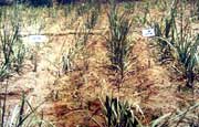
- The first
symptom of red rot in the field is discolouration
of the young leaves. The margins and tips of the leaves
wither and the leaves droop.
|
- The discolouration and withering continues from the tip
to the leaf base until the whole crown withers and the plant
dies, within 4 to 8 days.
- In a single stool, most of the stalks may wither almost simultaneously.
|
- As the disease
advances the entire stem rots and the central tissues
become pithy.
|
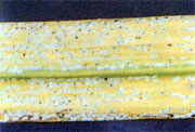
- The tissues are reddened throughout the basal portion,
especially the vascular bundles, which are intensely red;
there may be cross-wise white patches interrupting the reddened
tissues.
 |
- The internodes
may shrink and when such canes are split open large
cavities may be found in the centre and the pithy
tissues may appear brown.
- Often a profuse whitish growth of the fungal mycelium
may be found in the brown background of the host tissue.
- In some cases, black, minute, velvety bodies, representing
the acervuli of tire fungus, may also be seen
|
- Since reddening is a common symptom of other diseases
of sugarcane, the white patch symptom is an important diagnostic
characteristic of red rot.
- When a diseased plant is open, a characteristic becomes evident.
 |
- In the infected
plants the leaves may show symptoms in the form of
dark red lesions in the mid-rib, which may elongate,
turning blood-red with dark margins and later on with
straw-coloured centres.
- In the older lesions minute black dots, representing
the acervuli, can be seen. Often the infected leaves
may break at the lesions and hand down
|
Perfect stage : Physalospora tucumanensis
Spegazzini
Class : Ascomycetes
Order : Sphaeriales
Family : Mycosphaerellaceae
- The fungal mycelium is present inter-cellularly, mainly
in the parenchymatous cells of the path.
- The hyphae are thin, hyaline, septate, profusely branching
and contain oil droplets.
- The acervuli, which are characteristic of the genus, are formed
on the surface of the rind, in the leaf mid-rib and sometimes
in the pith region. They are minute, black and velvety.
- The compact hymenial layer produces the long, rigid, bristle-like
setae, which are septate, dark brown at the base and lighter
towards the tip.
- Inter-spaced between the setae are numerous club-shaped to
linear, hyaline, single-celled conidiophores, measuring approximately
20 X 8 m.
- The conidia, which are borne singly oil the conidiophores,
are single celled, thin walled, hyaline, falcate, granular
and guttulate, measuring 16-48 x 4-8m In artificial cultures
terminal and intercalary, thick-walled, greenish-black chlamydospores
are often produced.
- The conidia germinate readily to produce single germ tubes
at the tips of which an elliptical, ovoid or irregular, appresoria
are formed.
- The perfect stage of the fungus has been observed both in
nature and in culture by several authors.
Disease Cycle
- The planting material, viz., the setts, may harbour the
fungus and thus perptuate the disease from season to season.
- The fungus may also persist in the soil oil diseased clumps
and dry leaves left in the field after harvest.
- The primary infection, however, appears to be mainly from
infected setts.
- Secondary spread in the field may be through irrigation water,
cultivation tools anti implements and wind-borne inoculum.
- If the conidia settle on the leaves they may germinate and
invade the leaves through various types of wounds including
the splitting of the mid-rib so common in many varieties.
- Stem infection may take place through insect bores and root
primordia.
- The soil-borne fungus may also enter the healthy setts through
cut-ends, and cause early infection of the shoots.
- The prevalence of several pathogenic strains of the fungus
has been reported from many countries, including India.
- In general, light-coloured physiologic races sporulate readily
and are more pathogenic than the dark-coloured strains that
sporulate sparingly.
- Though the perfect stage of the fungus has been observed in
nature, the role of ascospores in the disease cycle is not
understood.
Control
- Red rot management in sugarcane has become an important
issue in all sugarcane areas. While early stage detection
may not be quite easy, during later stages, the cane breaks
down.
- The canes are to be split open lengthwise to see dull red
tissue, interrupted by white patches across the stalk. These
patches are characteristic of red rot of sugarcane. Monsoon
period enables faster disease spread and drying of the crop.
- The best remedy for avoiding this fungal disease is to cultivate
only resistant sugarcane varieties that have been released
for cultivation in different sugarcane growing states. Phytosanitation,
being the key to manage this disease, stringent domestic quarantine
measures to prevent movement of cane setts from endemic areas
to new areas has to be enforced by all concerned agencies
including sugar mills.
- In recent times, the disease has spread into many states.
- Following good cultural practices such as clearing fields
of excessive trash and ensuring efficient drainage.
- Healthy setts only are to be planted to avoid poor plant stand
due to rotting.
- Affected fields should be isolated through bunding to prevent
movement of water to adjacent fields.
- Rattooning of infected fields should be strictly avoided.
- Crop rotation in the affected fields could reduce disease
inoculum.
- Hot water treatment of setts before planting at 52 degree
C for 30 minutes is also recommended.
- Planting fungicide treated seed setts is to be followed as
a general practice to prevent introduction of the pathogen
into sugarcane fields.
- Chemical control: Sett treatment with 0.25% Thiophanate methyl,
a systemic fungicide will protect the setts against the primary
infection of red rot for 90 days.
- In addition to the various sanitary precautions mentioned
above, red rot may also be controlled by growing resistant
or tolerant varieties. Inter-generic and inter-specific crosses
with Saccharum spp., in the latter case using S.spopntaneum
and S. robustum with S. officinarum have yielded many economically
important cane varieties, some of which are highly resistant,
or at least tolerant, to red rot.
- Cultivation of resistant varieties: By far the most effective
measure for management of red rot is use of resistant varieties
for cultivation. Varieties viz. Co 8021, Co 85019, Co 86010
and Co 86032, Co86249 and Co 93009, Co 99004 and Co 99006
are tolerant/resistant to red rot in Tropical India. The
varieties Bo 91, Co89003, Co 98015, Co 99015, Co 99016,
Co S 96275, Co S 99259, Co Pant 90223 and Co Pant 94211
are resistant in SubTropical India.
Top
Red Stripe
Causal Organism : Xanthomonas rubrilineans
Class : Schizomycetes
Order : Pseudomonadales
Family : Pseudomonadaceae
- This is another bacterial disease of sugarcane which has
a world-wide distribution.
- It has been known since 1893 in Java and since 1903 in Australia,
but in India only since 1933, when it was reported by Desai,
and also McRae.
- In 1960 Rangaswami made detailed studies on the disease and
its causal agent.
Symptoms
- Red stripe is characterized by the appearance on the leaves
of chlorotic lesions carrying dark red stripes 0.5-1.0 mm
in breadth and several mm in length, either distributed all
over the blade, or concentrated in the middle
- Several of them may coalesce to cover large areas of the leaf
blade, and to cause wilting and drying of the leaves.
- Whitish flakes occur on the lower surface of the leaf, corresponding
to the red lesions on the upper surface.
- These flakes are the dry bacterial ooze. When young shoots
are affected, shoot or top rot may result.
- The growing points of the shoot are yellow and later reddish
with dark brown stripes on the shoots.
- The rotting may commence from the tip and spread downwards.
- If the affected plants are cut by splitting the shoot downwards,
dark red dis-colouration of the tissues may be seen.
- In the affected canes cavities may form in the pith region,
and the vascular bundles are distinct because of the dark
red dis-colouration.
- The diseased and rotting shoots can be easily pulled out and
separated from the plant.
Disease Cycle
- The disease spreads in the field by wind and rain, and
by cutting, as the basal stem from which the setts are taken
is mostly free from the bacterial infection.
- The bacterial ooze form the infected leaves and shoots dries
up to form a thin crust on the surface.
- When dry, the bacterial cells spread freely by wind.
- The cells falling on the host plants, enter them through natural
openings or wounds and establish themselves in the various
tissues, including the xylem.
- Infected parenchymatous cells may collapse and normal functioning
of the plant parts may fail.
- Several grasses, including ragi and bajra, have been reported
to be infected by the bacteria.
- These hosts may also play a role in the perpetuation and spread
of the pathogen.
Control
- This is a difficult disease of sugarcane to control.
- When-ever the disease is noticed, the affected plants should
be removed and burnt.
- Such systematic destruction of the affected plants reduces
the disease incidence.
- Growing resistant varieties is, however,the best method of
control.
Top
Sett rot or Pine Apple disease
Causal Organism
Ceralocystis paradoxa
Ceratostomella paradoxa
Class : Ascomycetes
Order: Sphaeriales
Family: Ceratostomataceae
- This disease of sugarcane is common in India and in most
of the sugarcane areas of the world including Hawaii, mainland
United States, Mexico, West Indies, parts of Africa, and South
America.
- In India it is found in all the sugarcane tracts.
Symptoms
 |
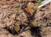
- The disease
primarily affects the sugarcane setts.
- When diseased setts are planted they may rot before
germination, or the shoots may die after reaching
a height of about 6-12 inches.
|
 |
- If infected
shoots survive, they are very much stunted and chlorotic.
- Eventually the leaves may wither and the shoots wilt.
- If the affected shoots and setts are examined the
central portion of the shoots will be seen discoloured
red and the contents of the sett rotting.
|
- A characteristic pineapple smell is associated with the
rotting and hence the name.
- The fungus produces both microconidia and macroconida.
- The microconidia are hyaline when young but become almost
black at maturity.
- They are thin walled aµm the chains of microconidia
are produced endogenousnd cylindrilorm, measuring 10-15 µm
x 3.5-5.0µm in the conidiophores and are pushed out
in succession.
- The conidiophores are linear, thin-walled, about 100µm
in length and are formed terminally on short basal cells.
- The macroconida are produced in chains of up to 20µm
short, lateral conidiophores, which measure 20-80 x 4 µm.
They are spherical, elliptical, truncale or sometimes pyriform
and are hyaline to olive-green or black, measuring 16-19 x
10-12 µm.
- The fungus produces the ascigerous of perfect stage in culture
media and at times on the host tissues.
- The perithecia are gregarious and flask shaped, with a long
narrow beak.
- The basal portion measures 200-350 µm in diameter and
the beak up to about 1200µm in length and 30-40µm
in diameter.
- The asci are clavate, and the ascospores are convex to elliptical
and measure 7-10 x 2.5-4 µm.
Disease Cycle
- The fungus is soil-borne, entering the setts through the
cut ends.
- Inside the sett it spreads rapidly through the parenchymatoous
tissues and causes sett rot.
- The buds on the setts may also be affected and secondary organisms
may hasten the decay.
- Occasionally the infected setts may also be responsible for
the spread of the pathogen.
- The micro and macroconidia may live saprophytically in soil
for several years and infect the setts when planted.
- Insects also play a part in the dissemination of the pathogen.
- The specific role of the ascigerous stage in perpetuating
the fungus is not under stood.
- The same fungus also infects banana, papaya, cocoa, coffee,
mango, pineapple, arecanut, coconut, palmhyra, date palm and
several other hosts.
Control
 |
- Healthy setts
should be obtained from disease-free field.
- They should be carefully selected and treated with
an organomereurial before planting, to protect the
cut-ends from invasion by the fungus.
|
- Pretreating the setts with hot water has been found to
stimulate germination of buds and hasten growth so as to help
the young plants to overcome the competition with the pathogen.
- A certain amount of resistance to the disease has been found
in a few sugarcane varieties in other countries, but field
sanitation practices combined with chemical pre-treatment
of the setts are quite effective in controlling the disease.
Top
Wilt
Causal Organism: Cephalosporium sacchari
Class: Deuteromycetes
Order: Moniliales
Family: Moniliacease
- This is one of the early known diseases of sugarcane in
India.
- It was first reported by Butler and Khan in 1913, from North
India.
- It has been reported to cause severe damage to sugarcane crops
in many parts of India. During 1965-1967 it caused severe
damage to sugarcane crop in the Deccan plateau
- Same disease has been reported from Mexico, Argentina, Barbados,
Columbia, Trinidad, Uganda, South Africa and United States
of America.
Symptoms
|
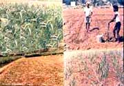
- The first
symptoms of the disease become apparent only when
the plant has grown for about 4-5 months.
- The canes show gradual withering.
|
- On examination of affected clumps , the pith will be seen
discoloured purple or dirty reef, with longitudinal streaks
- The leaves of affected clumps gradually turn yellow and dry
up.
- A characteristic disagreeable odour is also associated with
such diseased canes.
- A cottony white mycelium can also be seen in the pith region.
- Frequently this fungal disease is associated with a saprophytic
bacterial growth and often the bacteria are mistaken as causal
agents.
Disease Cycle
- The fungal mycelium is abundant in the infected canes.
- The hyphae are hyaline, thin walled and septate.
- They produce numerous microconidia on simple or branched,
lateral or terminal hyphae, but NO macroconidia are produced.
- This is an important character which is distint from that
of Fusarium. The conidia are oval to elliptical, and measure
4-12 x 2-3µm in size. They are mostly unicellular, but
the ones formed later in the advanced growth of the fungus
may be septate
- Conidia readily germinate to produce single germ tubes.
- The fungus is transmitted from place to place through the
infected seed setts.
- When the diseased setts are planted, the eyes may fail to
develop or often the shoots arising from the eyes may wilt,
due to the infection spreading to the shoots.
- Root formation in such setts may be very poor. The fungus
can also survive in soil as a saprophyte for 2-3 years.
- Near-neutral and alkaline soils are favoured by the fungus.
The perfect stage is not known.
Control
1. The disease is controlled by selecting seed setts from
disease free areas.
2. Alkaline soils may be avoided for growing the crop. The setts,
selected from disease free stalk, should be dipped in organomercurial
fungicide before planting.
3. Dipping the setts in 40 ppm of boron or manganese, or spraying
the plants with either of these minor elements reduces the
disease intensity.
4. The varieties Co-617 and B.P.-17 are more resistant than others;
hence they should be used when growing crop in the wilt-sick
soils.
Top
Grassy Shoot
- This disease was first observed in Maharashtra on sugarcane
variety Co. 419 in 1942.
- At present it is reported to be widespread in parts
of Andhra Pradesh, Telangana, Mysore, Bihar, U.P. and
Tamil Nadu.
- The disease is more severe on the ratoon crops than on the
first planted ones.
Symptoms
|
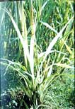
- The disease
is characterized by proliferation of vegetative buds
from the base of the cane giving rise to crowded bunch
of tillers bearing narrow leaves.
- The tillers bear pale yellow to completely chlorotic
leaves.
- Cane formation rarely takes place in affected clumps
and if formed the canes are thin with short internodes.
|
- The virus is readily transmitted by sap inoculation and
in the field it is transmitted through infected setts and
perpetuated through crop ratooning. The aphids are the vectors
for this disease.
- The same virus also infects jowar, Napier grass and Madras
grass.
Control
- The disease is controlled by eradication of diseased parts
as soon as symptoms are noticed
- Avoid selection of setts from diseased area
- pre-treating the healthy setts with hot water at 52°C
for 1 hour before planting
- Treating them with hot air at 54°C for 8 hours and spraying
twice a month with aphidicides.
Top
Gummosis or Gumming Disease
Causal Organism : Xanthomonas vasculorum
Class :Schizomycetes
Order :Pseudomonadales
Family :Pseudomonadaceae
- This is one of the oldest and most common diseases of
sugarcane known in many counties.
- The disease was first reported from Brazil in 1869, since
when it has been reported from Puerto Rico, West Indies, Columbia
and Australia.
- In India Rangaswami reported its occurrence in Tamil Nadu
State in 1960, and it is now known in other sugarcane areas
of South India.
Symptoms
- The disease is characterized by three distinct symptoms.
- On the leaves characteristic longitudinal streaks or stripes,
1/8 to ¼ inch in width and several inches in length
are found, these stripes are pale yellow in colour, later
turning brown.
- The affected canes are stunted with short internodes, giving
the plant a bushy appearance. When such canes are split open
or cut transversely, a dull yellow bacterial ooze comes from
the cut ends.
- In advanced cases, cavities may develop in the pith region.
- The fibro-vascular bundles are deep red and this is more intense
at the nodal regions of the stem.
- The causal bacterium is variously named by different authors,
but the above name is universally accepted at present.
- The bacterium is a hort rod, 1.0-11.5µm x 0.4-0.5µm,
motile by means of a single polar flagellum.
- Gram negative, non-spore forming, non-capsular and non-acid
fast.
- On beer extract agar it produces a yellow spreading and slimy
growth.
Disease Cycle
- The disease is primarily spread through the setts taken
from diseased plants.
- The secondary spread may be through agricultural implements,
including the cutting knife.
- In the field the infection may spread by wind and water, the
source of pathogen being the gummy bacterial ooze coming out
of diseased tissues.
- Certain insects, particularly flies, play a significant role
in transmitting the pathogen from place to place.
- The bacterium can survive in the insect's body for a long
time and in this way may be transmitted long distances.
- On entry into the host the bacterium reaches the vascular
tissues and becomes systemic.
- Besides sugarcane, several plant species falling under Graminae,
including maize, sorghum, Panicum spp. and some other grasses
have been found to be infected by this bacterium.
- These hosts may help in many ways the bacterial spread and
perpetuation.
Control
- As in other diseases of sugarcane, gummosis can be controlled
by selecting setts from disease-free plants, and adopting
strict field sanitation practices.
- In the Fiji Island the disease was completely eradicated by
destroying the diseased canes and growing resistant varieties.
- Studies on the availability of resistant stock among the sugarcane
varieties in India are needed.
Top
Foliar Diseases
- Pokkah Boeg caused by Gibberella fujikuroi (Saw.) Wollenw.
Wineland with its imperfect stage Fusarium moniliforme Sheldon.
- Downy mildew caused by Sclerospora sacchari Miyake.
- Eye spot caused by Helminthosporium sacchari (Breda de Haan)
Butl.
- Yellow spot caused by Cercospora Kopkei Krueg.
- Brown spot caused by Cercospora Longipes Butl.
- Leaf spot caused by Cercospora vaginae Krueg.
- Rust caused by Puccinia kuchnii (Kr.) Butl. and P.erianthi
Padwick & Khan.
- Ring spot caused by Letophaeria sacchari Breda de Haan.
- Leaf blotch or spot caused by Helminthosporium halodes Drechsler,
H. sacchari and H. tetramera McKinney.
- Root rot caused by Pythium spp.
- Smut caused by Sphacelotheca erianthi.
- Ergot and sugary disease caused by Claviceps spp.
- Root rot caused by Rhizoctonia solani Kuhn and Sclerotium
rolfsii Sacc. The symptoms of these diseases are similar to
the ones caused by species of the same genus on other hosts.
Top
Root-Knot Nematode
Casual Organism : Meloidogyne javanica
Class: Nematoda
Order : Tylenchida
Family: Heteroderidae
- Over 50 nematode species belonging to 20 different genera
are reported to infect sugarcane in various countries of the
world.
- At least 10 have been reported in India.
- Among these the Meloidogyme spp. are important.
- Sugarcane is among the various hosts affected by nematodes
in India.
- The crop is damaged by the root-knot nematodes, though several
other ecto-parasitic nematodes occur on this host.
- The occurrence of root-knot and other hosts was first established
by Rangaswami and his associates during 1958-1960.
Symptoms
- The diseased plants are chlorotic and stunted, and yellow
stripes show on the young leaves while older leaves appear
healthy.
- Crops at all stages of growth are affected and the symptoms
are more prominent during summer months.
- When the roots of affected plants are dug out and examined,
they are found to be knotted.
- The young, white roots show much less knotting than the older,
wiry ones.
- The knots are usually linear and are found more commonly towards
the root tips. They are about 5-8 mm in thickness.
- The nematodes are separated from the infected tissues by the
Baermann Funnel technique and examined under the microscope.
- The females measure 400-460mm in length and 45-50mm at the
thickest point, oesophagus 40-45mm and tail 30-35mm.
- The males measures 420-460mm in length, 20-25mm in width at
the thickest point and oesophagus 55-60mm and tail 30-35mm.
- The cyst is pear shaped, measuring 600-650mm x 380-400mm with
a prominent beak.
- The other species of Meloidogyne reported on sugarcane are
M. arenaria Chitwood and M. incognita Chitwood, both of lesser
importance than M. javanica.
Disease Cycle
- The nematode eggs persist in soil in cyst form and when
the susceptible host is planted, the cyst give rise to the
larval forms which invade the root and form the galls.
- There are reports on the exudation of specific chemical substances
by the various hosts with either attract or repulse the nematode
larvae.
- Once the larvae are inside the host tissue, they feed through
the vascular bundles and complete the life cycle.
- They produce the cysts, which protrude from the host roots
and remain there until harvest.
- The production of growth regulators such as indole acetic
acid in the host tissue is reported to be correlated with
the growth promoting and nodule forming effect of the nematodes,
as found by Rangaswami and his associates.
- There are over one hundred plant species, belonging to diversified
families, which are host plants for M. javanica. No doubt
these host plants, particularly the weeds present in sugarcane
fields, and the crops used in rotation with sugarcane, play
significant roles in the perpetuation and inoculum build-up
of the nematode.
Control
- The root-knot nematode can be checked by fumigating the
soil with a nematicide such as Nemagon or D.D.T
- The soil must be opened up soon after crop harvest.
- The chemical fumigant should be injected into the soil as
per the directions given by the manufacturer after which the
soil is kept covered with a tarpaulin or plastic sheets for
a week or two, if necessary.
- The fumes permeate the soil, killing the nematode larvae and
cysts, and other organisms as well.
- The soil is safe for planting in 2-3 weeks after fumigation.
- In some countries crop rotation with marigold reduces the
nematode population.
- The effect of such plants or their plant roots exudates on
the nematode population, and the inter-relationships of other
soil microbial populations with the nematode, have to be investigated
in detail to understand this complex phenomena has to evolve
effective control measures.
- Certain nematophagous fungi like Catenaria vermicola, Arthrobotrys
conoides Drech. and Dactylella deodycoides Drech. are present
in soils, and this might help in reducing the nematode population.
- Besides Meloidogyne javanica and other species of Meloidogyne
which cause root-knot symptoms, Pratylenchus sp., Paratylenchus
macrophallus deMan, Trichodorus sp., Rotylenchus sp., Tylenchorhynchus
sp, Xiphinema sp., Rotylenchus sp. and Hoplolaimus coronatus
Cobb have been reported by Kishan Singh on sugarcane in some
parts of India.
- More detailed studies are needed on the disease symptoms,
life cycle of the nematodes, nature and estimation of damage
and control measures.
- A phanerogamous parasite affecting the roots of sugarcane
has also been reported from some parts of India.
- It is identified as Striga euphrasioides Benth.
- This is a partial root parasite, growing up from the roots
to form leafy shoots.
- The parasite can synthesize carbohydrates through the green
chlorophyll pigments in the leaves but for its other nutrients
it depends on the host root.
- It is usually controlled by pulling out the shoots before
flowering and seed set.
- Spraying weedicides like 2-4, D will kill the parasite, without
affecting the sugarcane plant appreciably.
Top
Ratoon Stunting
Symptoms
|
|
- This is a
virus diseases of sugarcane found in some parts of
India in recent years.
- The affected plants are stunted, the stunting being
most severe in stubble and ratoon crops.
- The setts taken from diseased plants germinate poorly
and the few shoots that emerge grow very slowly.
- The virus infects many hosts falling under Graminae.
- It is sap-transmissible and no insect vector has been
found.
|
Control
- The disease spreads mainly through planting material,
setts should be selected from healthy plants.
- Treating the seets in hot water at 500C for about 2 hours
gives 100 per cent control and has become a regular practice
in many countries.
Top
Mosaic
Symptoms
|
|
- This disease
was first observed in Java in 1892 and since then
it has been reported from almost every sugarcane growing
tract of the world.
- The disease carries different names in different countries
but the most common name is 'sugarcane mosaic.'
- During 1919-1920 it caused devastating damage to the
sugar industry in Puerto Rico and Cuba, and this led
to concentrated studies to establish the causal agent,
which was uncertain until about 1920.
|
- In India the disease was identified during the early thirties
of this century and has been reported since then from almost
all the sugarcane tracts of the country.
- The intensity of the disease and the loss in crop yield vary
considerably, depending upon the host variety and climatic
conditions.
Causal Organism
- Sugarcane Mosaic Virus (Marmor sacchari).
- The virus is present in abundance in the chlorotic lesions
of the affected leaves.
- It causes varying degrees of destruction of the chlorophyll,
and hence the chlorotic symptom.
- The virus has been purified and studied in detail for its
physical and chemical properties.
- Purified virus particles are rods, measuring the range of
650-770 x 12-15 mm.
- Several strains of the virus, designated as A, B, C, D, E,
F and G and a few sub-strains have been differentiated, based
on their physical properties and virulence.
Disease Cycle
- The disease spreads readily through infected canes used
as seed setts. It is also transmitted from plant to plant
by the aphids Rhopalosiphum maidis (fitch) and Hysteroneura
setariae Thos.) and by the green bug Toxoptera graminum (Rond.)
- The virus is a non-persistent type in its vectors.
- In India R. maidis is the main vector; thogh T. graminum (Schizaphis
graminum) was demonstrated to be a vector of the virus in
experimental studies.
- The virus is also sap-transmissible.
- Once the virus is inside the susceptible host tissues, it
becomes systemic.
- The incubation period varies from 7 to 20 days, depending
upon the host variety and virus strain.
- The virus spreads, both from the leaves to the stem and from
the stem to the leaves, and to other shoots in the clump.
- Depending upon the climatic and other environmental conditions
and host-pathogen relationships, the symptoms may be prominent
or masked.
- In some cases plants have recovered from early infections,
due to changed environment and altered physiological condition
of the host plant.
- From such plants, healthy shoots may arise from the clump
and the newly formed leaves may have no mosaic symptoms.
- In such cases the damage to the crop in reduced yield and
sugar content is less.
- Besides sugarcane this virus infected several other cultivated
plants, including sorghum, maize and some wild grasses
Control
- In the early years of this disease attempts were made
in Java and other countries to completely eradicate it by
cutting and burning the diseased plants.
- Later it was realised this not only was not easy to do, but
also it was necessary because of the availability of resistant
varieties.
- Roguing infected plants and chemical sprays to kill the insect
vectors are also advocated.
- However, the most practical method of controlling the disease
is to grow mosaic-resistant or, at least, tolerant varieties.
- Introduction of mosaic-susceptible varieties during earlier
years, produced at the Sugarcane Research Institute, Coimbatore,
in other areas of the world often resulted in devastating
damage to the crop.
- This emphasized the need to breed mosaic-resistant varieties.
- At present the newer varieties are invariably tested for resistance
to mosaic and other severe disease, before they are released
for large-scale cultivation.
- Saccharum spontaneum L. and S. barberi Jesweit carry resistance
to mosaic.
- Most of the popular sugarcane varieties under cultivation
in India are resistant or tolerant to the disease.
- The occurrence of another virus disease, Ring mosaic, on sugarcane
in India was also reported recently.
- It causes ellipitic rings with well developed yellowish or
dark green centres.
Top
|